Cytos™ Protein Catalog
Further validation detail for every target in our fixed panels — organized by panel, so you can judge fit before you order.

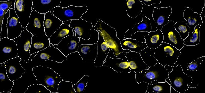
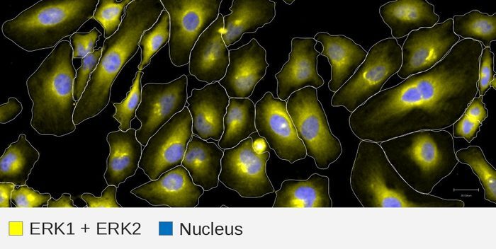
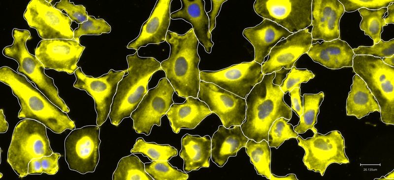
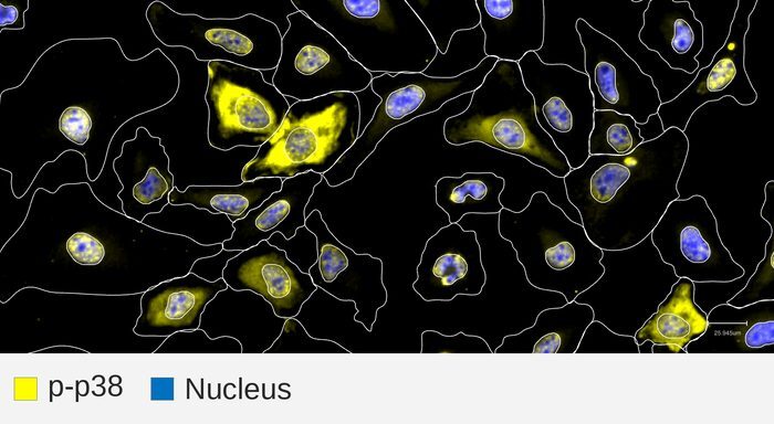
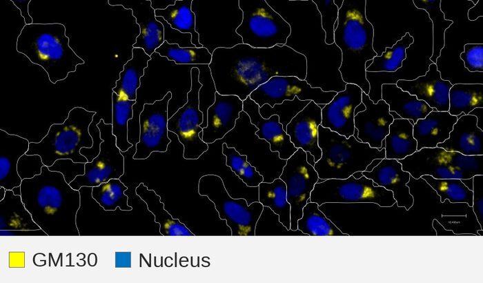
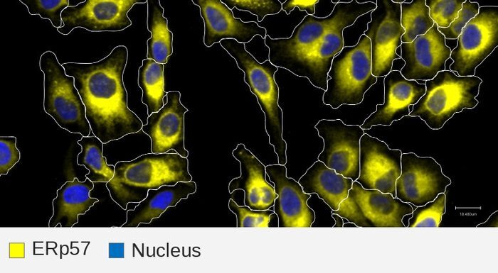
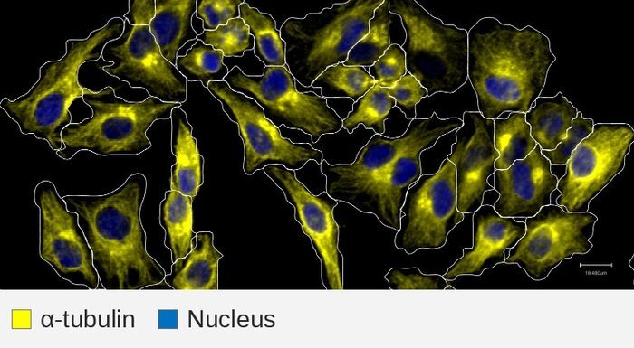
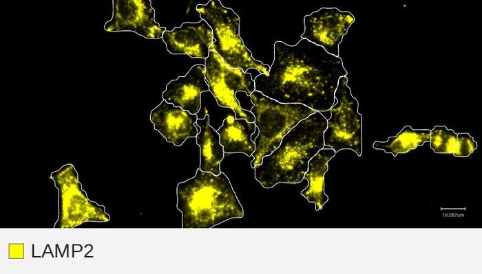
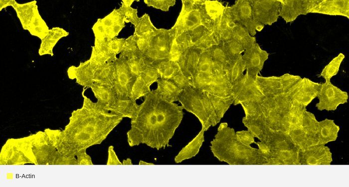
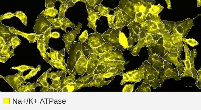
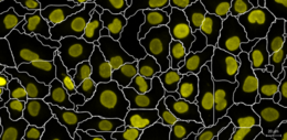
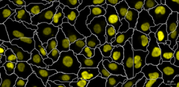
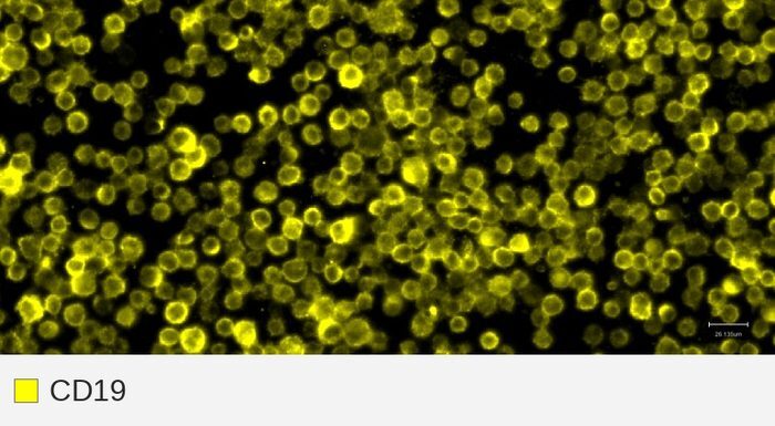
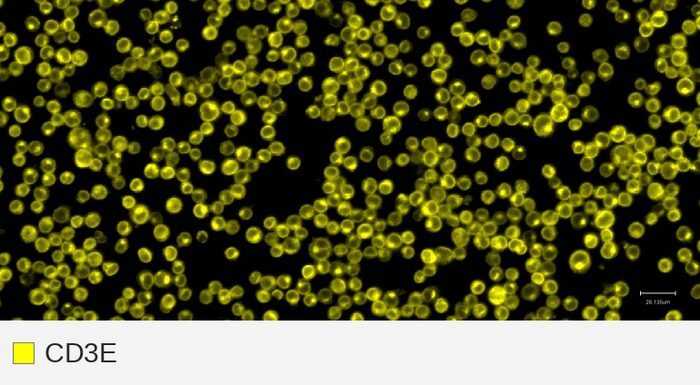
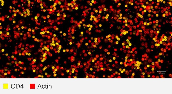
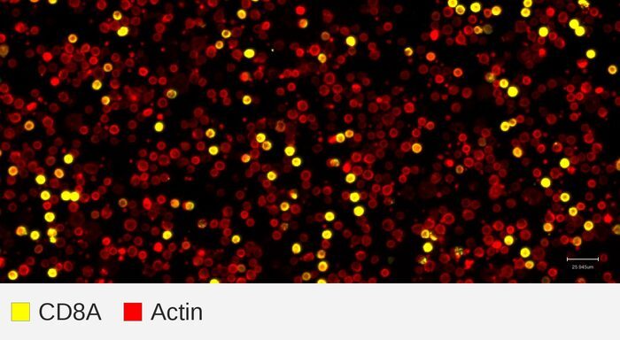
ⓘ Fixed panel targets. These proteins are part of our supported panels. We provide validation information for each — click any target below for its report card. Targets are labeled in yellow and nucleus in blue.
Expanded Morphology Panel
Adds extra organelle and structural stains on top of the core Cell Paint markers, extending coverage to the Golgi, ER, microtubules, and lysosomes.
Neuro Morphology Panel
Improves Cell Paint staining in neuronal and glial cell types (iPSC-derived neurons, astrocytes, microglia, SH-SY5Y) where the core Cell Paint markers may not perform as well. These markers can also be used to re-segment cells when quality is better than core Cell Paint.
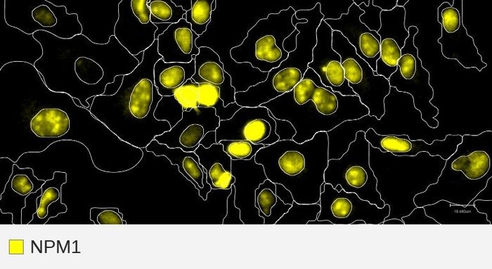
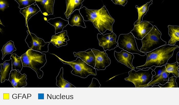
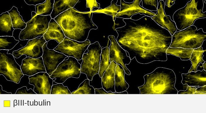
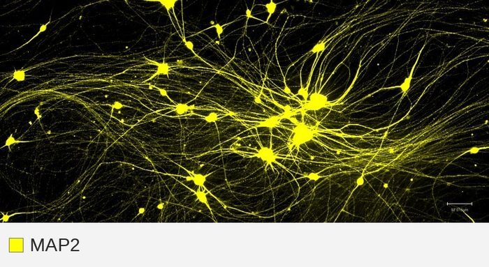
Alternative Morphology Panel
A backup set of the same core markers (membrane, actin, nucleus, nuclear envelope) for cells cultured on laminin or matrigel and/or non-standard or unvalidated cell types, in case the core Cell Paint stain may not perform as well. These markers can also be used to re-segment cells when quality is better than core Cell Paint. See our validated cell types for guidance.
Immune Subtyping Panel
Core B and T cell lineage and subset markers.
Cell Signaling Panel
Canonical pathway-activity markers for tracking signaling state — MAPK/ERK activation and stress response — across conditions.