Protein Overview
p38 MAPK is a stress-activated kinase of the SAPK/MAPK superfamily. Dual phosphorylation at Thr180/Tyr182 in its activation loop is mediated by the upstream MAP2Ks MKK3 and MKK6, marking full activation; active p38 phosphorylates MAPKAPK2/3, transcription factors (ATF2, MEF2C), and other stress-response effectors.
Expected Localization
Predominantly cytoplasmic in unstimulated cells; upon activation it partially translocates to the nucleus to phosphorylate nuclear substrates while retaining a cytoplasmic pool.
IF of stimulated cells shows increased nuclear staining intensity superimposed on cytoplasmic signal.
Expected Expression Pattern
Broadly expressed across most mammalian cell types, including monocytes/macrophages, epithelial cells, fibroblasts, and cardiomyocytes. Phospho-Thr180/Tyr182 signal is low at baseline and strongly induced by UV irradiation, osmotic shock, heat shock, oxidative stress, and pro-inflammatory cytokines (TNF-α, IL-1, LPS).
Relevant Pathways
- p38 MAPK stress-activated signaling cascade
- MKK3/MKK6 upstream kinase activation
- Inflammatory cytokine signaling (TNF-α, IL-1, LPS/TLR)
- Downstream MAPKAPK2/3-HSP27 signaling axis
- Apoptosis and cell-cycle checkpoint regulation under stress
Clone Information
Antibody Species: Rabbit
Cell Type Compatibility: Validated in human cells only using Cytos™ kits.
For more details on the clone and reactivity with other species, visit ABclonal and search for the clone catalog # AP1508BCS.
Qualitative Results
Localization Assessment
Phospho-p38 (T180) protein panel demonstrates strong localization to nuclear speckles and the cytosol. The observed localization of Phospho-p38 (T180) protein is consistent with published data. Localization is dependent on cell stress level.
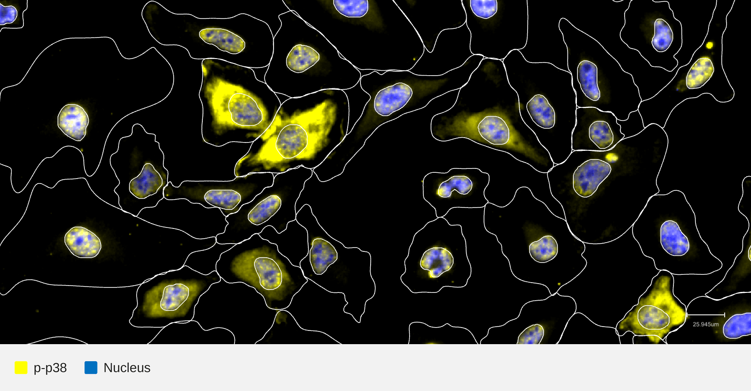
| HeLa TNFα 30 min |
HeLa cells were cultured in a 12-well format Cytos slide kit overnight, treated with 100 ng/mL TNFα for 30 min, fixed with 4% formaldehyde for 30 minutes, permeabilized using 70% ethanol for 10 minutes, and then treated with Lambda Phosphatase (LPP, NEB P0753L) at 30 °C for 1 hour. After that, cells were incubated with probe made with biotinylated rabbit monoclonal anti-Phospho-p38 (T180) antibody for 1 hour at room temperature. Negative control probe based on Biotin Rabbit Isotype Control (ABclonal AC042BC) was included to assess background signal and potential crosstalk, with no detectable crosstalk observed. The slide kit was then assembled into a flow cell and loaded onto an AVITI24™ along with the Cytos Protein Cartridge for a multiomics cell screening run. Phospho-p38 (T180) demonstrated strong cell status specific localization and high signal-to-noise ratio, with no detectable probe crosstalk. These results support high confidence in target-specific detection and validate this antibody as a passing clone.
Quantitative Results
| Signal-to-noise ratio Acceptance criteria: > 2x background |
Pass |
- 6.5 (HeLa, TNFα 30 min, −LPP)
Indicates the fold brightness of the target signal as measured by the Mean Intensity Above (Otsu-defined) Threshold (IAT), over the background (a channel-matched isotype control) in the same cell. The ratio of 6.46 in TNFα-stimulated HeLa cells without phosphatase treatment is over 3 times the acceptance threshold.
|
Panel-level run-to-run reproducibility |
Pass |
For each imaging panel, we assess reproducibility by comparing the intensity signal a given antibody target produces across independent replicate runs. Within each run, we measure the average per-well fluorescence intensity for every target in the panel. We then plot each target's intensity from one run against the matching target's intensity from another run of the same layout, and calculate the Pearson correlation coefficient (r) between those two sets of measurements. Squaring that value gives the R² (coefficient of determination), which reports what fraction of the variation in one run's signal is explained by the other run's signal.
Disclaimer: This information is provided to help assess whether the target suits your experiment. It reflects internal testing conditions and does not guarantee performance in your application. Performance in other cell types and species such as mouse or rat may require additional validation using Cytos kits as performance is not guaranteed.
ELEMENT BIOSCIENCES For Research Use Only. Not for Use in diagnostic procedures.