Highlights
- Direct in Sample Sequencing (DISS) on the AVITI24™ system with Cytos chemistry reads single guide RNA (sgRNA) sequences directly from cells, with no poly-A capture required
- Link sgRNA assignment with cellular morphology along with 3’ transcriptome data and immunofluorescence-based protein detection, all at a subcellular resolution
- Choose good sgRNA scaffold design and apply proper multiplicity of infection (MOI) and selection to increase CRISPR effect and assignment rate of cells with guide detected
Introduction
Pooled CRISPR screens enable systematic interrogation to support research into gene function, disease mechanisms, and targeted discovery. Traditional methods for phenotypic assessment of pooled CRISPR perturbations leverage imaging-based readouts for morphological profiling or single-cell transcriptomics to resolve gene expression differences between perturbed cells. These approaches generally capture one phenotypic modality and often require trade-offs between screening scale, cellular coverage, and cost.
Linking phenotype to perturbation requires reliable guide detection. Because sgRNAs are not naturally polyadenylated, transcriptomic methods need specialized capture strategies, while optical methods may require integrated custom capture sequences or optimized probes for hybridization or in situ sequencing-based detection. Depending on the approach, these requirements can limit library compatibility or add custom probe design complexity.
The Cytos Optical Pooled Screening (OPS) Kit utilizes Direct In Sample Sequencing (DISS) on the AVITI24 system to sequence sgRNA in cells with morphology metrics. The Cytos Multimodal Pooled Screening (MPS) Kit extends the same workflow with 3’ transcriptome data, while the Cytos Protein Kit enables immunofluorescence-style protein detection. A predesigned quality control panel targeting two highly expressed transcripts, ACTB and GAPDH, with 4 targets each, serves as a control for guide detection, and is included with each OPS and MPS kit. These kits provide multimodal data from a single pooled screen that delivers guide identity, morphology, transcriptome, and protein abundance and localization.
Cytos CRISPR Screening Workflow
Figure 1 shows the DISS workflow for targeted RNA. In this workflow, a custom, user-designed reverse transcription (RT) primer binds to the 3’ flanking region of a target RNA sequence. Reverse transcription generates cDNA that contains the target sequence, which is then amplified with rolling circle amplification (RCA) and sequenced using Avidite Base Chemistry™ with a user-designed sequencing primer.
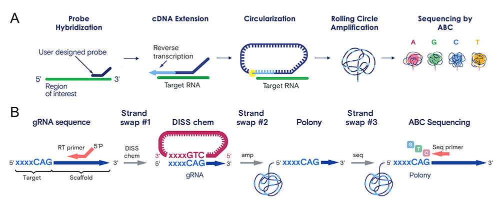
Figure 1. (A) Targeted Direct in Sample Sequencing (DISS) workflow and (B) DISS chemistry steps showing strand swaps.
The ElemBio Cloud Custom Designer™ automates primer design with user-specified guide scaffold sequences, and primers are ordered directly from third party oligo vendors. The custom RT primer and sequencing primers are added to the 3’ Transcriptome Cartridge in a Cytos OPS or MPS Kit during run set-up. You can learn more in the Custom Primer Design section of the Cytos Online Help. In CRISPR screening applications, the Cytos assay reads expressed sgRNA spacer sequences within each cell. Automated onboard assignment of sequences to sgRNAs and sgRNAs to cells is performed for experiments that include a Target Cell Assignment Manifest. A Confidence Score is calculated to represent cells that are confidently assigned to a single sgRNA target. By default, cells with more than one guide are assigned to a dominant spacer if > 50% of guides are assigned to that known spacer. For CRISPR systems with multiple guides per cell, the criteria for cell assignment should be adjusted accordingly.
Scaffold Design and sgRNA Detection
Previous studies1,2 have demonstrated that modifying the sgRNA scaffold sequence and structure increases intracellular sgRNA levels and enhances gene knockout efficiency, which can subsequently improve guide detection with Cytos. The original sgRNA scaffold design in these studies contained a run of four consecutive uridines, which functioned as an RNA polymerase III transcriptional terminator that can cause premature termination of sgRNA transcription. In the optimized scaffold design, mutating the U-A base pair at position 4 of this run to a C-G pair significantly increased sgRNA levels. In addition, extending the Cas9-binding hairpin by 5 base pairs improved sgRNA-Cas9 assembly, further increasing sgRNA levels in cells. Combining both modifications increased sgRNA levels approximately five- to six-fold relative to the original scaffold and yielded the highest knockout efficiency.
These scaffold design improvements showed higher guide RNA counts and assignment in Cytos experiments, and are recommended for use with Cytos runs. Table 1 and Figure 2 show six different guide libraries expressing sgRNAs under various constructs that were assayed on a 12-well Cytos Slide Kit (2 wells per cell type + construct). The libraries with the optimized sgRNA construct yielded approximately 20–40 counts per cell, while the libraries without this scaffold typically yielded fewer than 15 counts per cell (Table 1, Figure 2). For assistance implementing these design modifications, contact Element Biosciences™ Support. Note that multiple factors such as the MOI, passage number, cell type, and antibiotics selection mechanism determine the number of unique guides expressed within a cell and the frequency of cells without guide expression.
|
Library |
Vector Style |
sgRNA Promoter |
gRNA scaffold |
Cell Type |
% Cells Assigned |
Mean sgRNA counts/cell |
Mean sgRNA counts/mm2 |
|
1 |
CROP-seq-opti |
U6 |
Optimized |
U2OS | 96.6% | 42.8 | 54,155 |
|
2 |
PerturbView |
U6 |
Optimized |
MCF7 | 70.6% | 22.1 | 41,554 |
|
3 |
Cellecta pRSGUPS2 |
U6 |
Optimized |
A549 | 86.4% | 20.7 | 12,505 |
|
4 |
lentiGuide-Puro |
U6 |
Original |
U2OS | 65.2% | 12.1 | 10,494 |
|
5 |
pLentiCRISPRv2 |
U6 |
Original |
A549 |
52.8% |
9.8 |
8,001 |
|
6 |
pLentiCRISPRv2 |
U6-T7 Hybrid |
Original |
HeLa |
14.5% |
1.4 |
1,510 |
Table 1. Six guide libraries (three with optimized scaffold and three with original scaffold) assayed with Cytos Optical Pooled Screening Kit and Cytos Slide Kit-12-well. Percent of cells assigned (with Confidence Score > 0.5), mean sgRNA counts per cell, and mean sgRNA counts per mm2 are listed in the table. The number of assigned sgRNA counts per cell is reflective of the total sgRNA detection level in the cells.
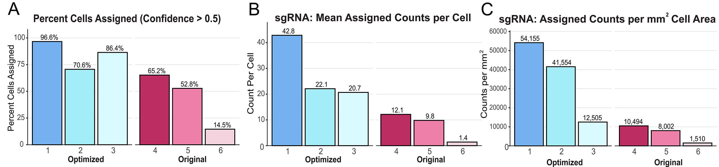
Figure 2. Graphical representation of Table 1. (A) Percent of cells assigned with confidence score > 0.5, (B) Mean assigned counts per cell, and (C) Assigned Counts.
Practical Considerations
When designing CRISPR screening experiments, consider the number of cells needed per condition. Use a pilot experiment or approximate ~50% guide assignment as a starting point for experimental design.
For example, a Cytos run with a 1-well flow cell recovering ~200,000 assigned cells could support up to 500 conditions if 400 cells are required per condition to saturate the transcriptome space (200,000/400). Alternatively, a 2,000 gene perturbation screen at 400 cells/perturbation would require 800,000 total assigned cells. A typical run with a 1-well A549 flow cell might recover ~400,000 cells, so at ~50% assignment, this experiment would require roughly 4 flow cells (800,000/200,000), though the actual number will vary based on cell line, flow cell configuration, and coverage variability. These calculations are illustrative; the ideal numbers are best determined by the desired statistical criteria specific to the experiment. As shown in Figure 3, an A549 cell line infected with a 28-guide sgRNA library (six target genes and one non-targeting control, four guides each) displayed uneven cell counts per gene target, reflecting expected differences in cloning efficiency, MOI, and the impacts of sgRNA perturbations on cells.
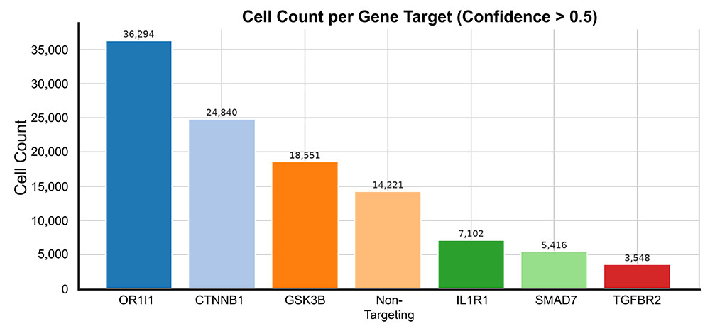
Figure 3. Total cells assigned across 6 wells of a 12-well flow cell, shown as cell count assigned per gene target for an A549 cell line tested with Cytos for CRISPR screening. The cell line was infected with a 28-sgRNA library targeting 6 unique genes and 1 non-targeting control, each with 4 unique guides. The sum of assigned cells across 6 wells was 109,972.
Other parameters, such as the use of fluorescent reporters for selection in CRISPR screening experiments, should be addressed before a Cytos run, particularly for experiments that rely on protein or morphological data. See the Fluorescent Reporters Knowledge Base article for more details.
Summary
The Cytos Optical and Multimodal Pooled Screening workflows simplify pooled CRISPR screening by sequencing sgRNA spacer sequences directly from fixed cells using Direct In Sample Sequencing. For rich, multimodal phenotyping, perturbation identity can be linked to morphology, 3' transcriptome, and immunofluorescence-based protein detection all at subcellular resolution. See the Cytos MPS preprint and Protein Detection Technical Note for more details. Together, simplified and scalable primer design, automated workflow and analysis, and multimodal read-outs enable high-resolution CRISPR screening at any scale, from focused campaigns to genome-wide screens across multiple flow cells.
References
- Dang, Y. et al. Optimizing sgRNA structure to improve CRISPR-Cas9 knockout efficiency. Genome Biol. 16, 280 (2015).
- Chen, B. et al. Dynamic Imaging of Genomic Loci in Living Human Cells by an Optimized CRISPR/Cas System. Cell 155, 1479–1491 (2013).
For Research Use Only. Not for use in diagnostic procedures.
LT-00084 | Rev A | October 2026