Question
How can I improve my cell culture techniques for optimal Cytos cell screening images on the AVITI24™ system?
Answer
Cytos cell screening images are influenced by a variety of lab techniques during the cell seeding, fixation, and permeabilization protocols in the Cytos workflow. Revisit pipette handling, seeding, distribution, fixation, and wash techniques when troubleshooting suboptimal results. See the Cytos Sample Preparation and Optimization User Guide for detailed protocols. Additionally, images are provided in the Troubleshooting section of the Cytos Sample Preparation and Optimization User Guide that can help identify potential issues.
Symptoms
Poor cell seeding and pipetting techniques can cause the following phenotypes in cell screening images:
- Ring-shaped area of cell loss
- Cell loss in well corners
- Cell loss in the middle of a well
- Scratch marks across a well or random cell loss
Considerations
To ensure the best cell seeding results, practice pipetting techniques such as slow pipetting and proper dispersion. See the following resources for detailed instructions, photos, and videos:
- Cytos Sample Preparation and Optimization Guide
- What are some best practices for culturing adherent and suspension cells with the 12-Well Cytos Slide Kits on the AVITI24 system?
- Can I use the same steps for cell culture and sample preparation in the 48-Well and 1-Well Cytos Slide Kits?
- Cell Culture Protocols
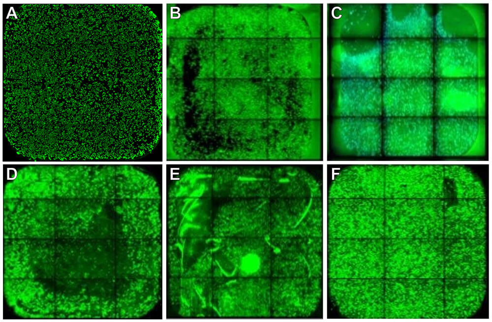
Figure 1A shows an example of good sample preparation techniques in a 12-Well Cytos Slide Kit. Figure 1B-F shows examples of poor seeding and pipetting techniques in a 12-Well Cytos Slide Kit. For details and recommendations, see the Troubleshooting section of the Cytos Sample Preparation and Optimization User Guide. Some of these phenotypes are also observable with brightfield microscopy before a run. For examples of brightfield images of various cell lines at different confluencies, see their associated Cell Culture Protocols and Cell Culture Hub.
- Successful preparation: even distribution of cells across the well without cell clumping. Confluency 50–70% (optimal confluency depends on cell type), absence of nicks or scratches.
- Ring-shaped area of cell loss: can be caused by circular movement of the pipette during seeding or of the slide kit during distribution. Seed cells in the center of the well without moving the pipette. Use a forward-and-back then side-to-side motion for uniform cell distribution.
- Cell loss in corner: can be caused by bubbles when applying a surface coating, seeding cells, or contact with the slide when pipetting. Pipette slowly to prevent bubbles when dispensing cells and check if a centrifugation step is required for your cell type or slide kit.
- Cell loss in middle: can be caused by adding liquid into the middle of the well after seeding. Pipette slowly and against the well wall after cells are seeded.
- Scratch marks on surface: caused by pipette tip contact with the surface after surface coating. Avoid touching the slide surface with the pipette tip after surface coating.
- Random cell loss: caused by pipette tip contact with the surface. Avoid touching the slide surface with the pipette tip.
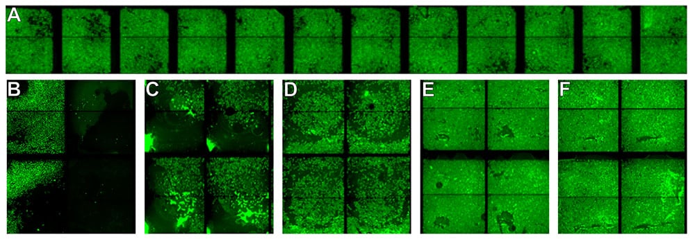
Figure 2A shows an example of good sample preparation techniques in a 48-Well Cytos Slide Kit. Figure 2B-F shows examples of poor seeding and pipetting techniques in a 48-Well Cytos Slide Kit.
- Successful preparation: even distribution of cells across the wells without clumping, minimal nicks or scratches.
- Cell loss in corners: can be due to bubbles. Ensure all liquid makes contact with the slide when seeding cells and applying surface coatings. Note that 48-Well Cytos Slide Kits require a centrifugation step to remove bubbles, even for adherent cells. This is because bubbles are more likely to form in the 48-Well Cytos Slide Kits when liquid is added due to their design.
- Cell detachment: can be caused by liquid impact. Pipette slowly and against the well wall.
- Ring-shaped area of cell loss: can be caused by circular movement during distribution. Ensure an even distribution of cells using a forward-and-back then side-to-side motion.
- Random cell loss: can be caused by pipette tip contact with the surface. Avoid touching the slide surface with the pipette tip.
- Scratch marks on surface: caused by pipette tip contact with the surface. Avoid touching the slide surface with the pipette tip.