Background
This study demonstrates the application of Direct In Sample Sequencing (DISS) in a pooled CRISPR screen of guide-expressing A549 lung epithelial cells (Cellecta cell line). Leveraging DISS, we directly sequenced perturbation identities and the 3’ transcriptome within the same cells used for imaging, linking CRISPR-induced changes to phenotypic, transcriptomic, and protein-level readouts. This approach enables high-content, multimodal analysis of how inflammation, Wnt, and TGF pathway perturbations shape cellular responses under a subtle TNFα stimulation.
Experimental Design & Methods
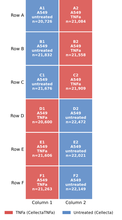
A549 Cas9-expressing cells were transfected with a 28-guide CRISPR library targeting CTNNB1, GSK3B, IL1R1, SMAD7, and TGFBR2, along with OR1I1 (olfactory receptor; no expected phenotype in A549) and non-targeting controls. Guide sequencing, 3’ transcriptome, cell paint, and 4-plex protein detection was performed using Cytos™ Multimodal Pooled Screening and the Cytos Protein Cell Signaling Panel. Cells were seeded in a 12-well Cytos Slide Kit and profiled at baseline and following 30 mins TNFα stimulation, with 6 replicates per condition resulting 258,277 total cells profiled. In total, 87% of cells were assigned to a gRNA with 1,600-1,800 RNA counts/cell on average.
Data highlights
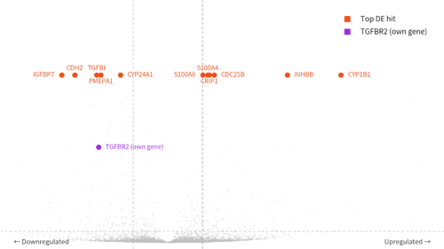 |
TGFBR2 knockout downregulates TGF-β targetsTGFBR2-KO cells show loss of the receptor transcript itself, confirming on-target editing, alongside reduced expression of canonical SMAD-dependent targets PMEPA1, TGFBI, CDH2, and IGFBP7—the expected direction of effect for disrupting the TGF-β axis. It confirms the pooled knockout phenotype is captured cleanly at the single-cell transcriptomic level.
|
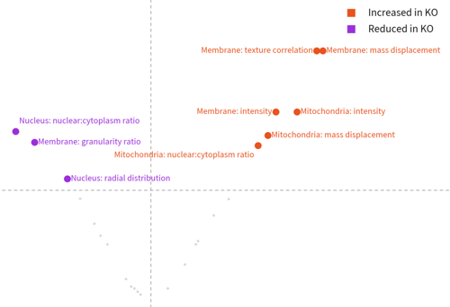 |
TGFBR2 loss produces a matching morphological phenotypeSignificant features reflect organelle/membrane redistribution toward a clustered, perinuclear state consistent with TGFBR2's known role in cytoskeletal organization, and corroborated by this run's own RNA data showing shifts in cytoskeletal/adhesion genes (CDH2, ITGAV, TGFBI).
|
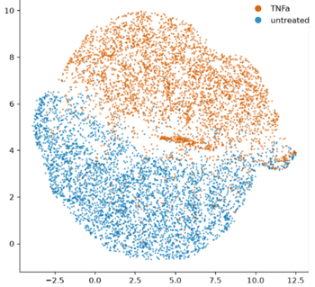 |
TNFα treatment resolves protein-level state within 30 minutesUsing the Cytos Protein Cell Signaling panel, which includes Phospho-p38, Phospho-ERK1/2, Phospho-HSP27, and ERK1/2, treated and untreated cells resolved into two largely distinct populations by UMAP after just 30 minutes. This clean separation reflects the fast kinetics of post-translational signaling: phosphorylation-driven stress responses are already underway before the transcriptional program has fully caught up, and this assay captures both layers—protein and RNA—from the same experiment. |
Download the full dataset
Explore data from a Cytos Multimodal Pooled Screen on AVITI24™.