Protein Overview
Alpha-tubulin is a core structural component of microtubules. Alpha- and beta-tubulin heterodimers polymerize to form microtubules that maintain cell shape, organize intracellular architecture, support organelle and vesicle transport, form cilia and flagella, and assemble the mitotic spindle required for chromosome segregation.
Expected Localization
Cytoplasmic microtubule network, centrosomes, mitotic spindle, midbody, and microtubule-based cellular projections.
Interphase cells typically show a filamentous network radiating through the cytoplasm, while mitotic cells display prominent spindle structures.
Expected Expression Pattern
Ubiquitously and abundantly expressed in most eukaryotic cells. The organization of the staining pattern changes markedly with cell-cycle stage, cell morphology, differentiation, and exposure to microtubule-stabilizing or -destabilizing agents.
Relevant Pathways
- Microtubule cytoskeleton organization
- Mitotic spindle assembly and chromosome segregation
- Intracellular vesicle and organelle transport
- Cell polarity and migration
- Cilium and flagellum organization
Clone Information
Antibody Species: Rabbit
Cell Type Compatibility: Validated in human cells only using Cytos™ kits.
For more details on the clone and reactivity with other species, visit ABclonal and search for the clone catalog # A6830BCS.
Qualitative Results
Localization Assessment
α-tubulin protein panel demonstrates strong localization to microtubules. The observed localization of α-tubulin protein is consistent with published data.
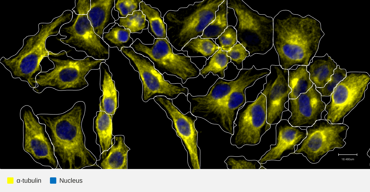
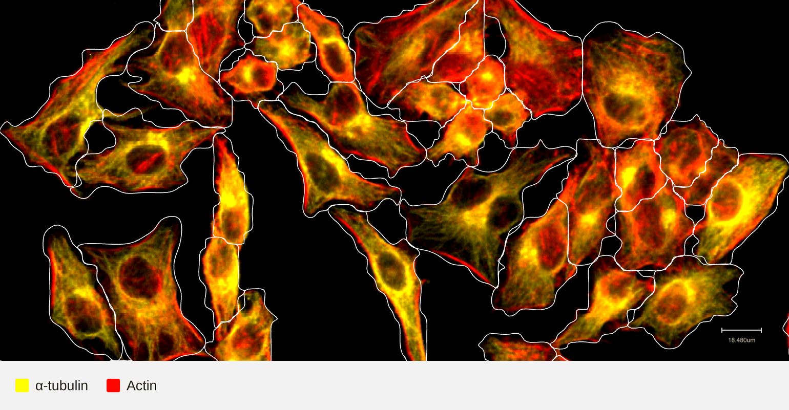
| HeLa | HeLa |
HeLa cells were cultured in a 12-well format Cytos slide kit overnight then fixed with 4% formaldehyde for 30 minutes, permeabilized using 70% ethanol for 10 minutes. The slide kit was then assembled into a flow cell and loaded onto an AVITI24™ along with the Cytos Protein Cartridge for a multiomics cell screening run. α-tubulin demonstrated strong microtubule localization and high signal-to-noise ratio, with no detectable probe crosstalk. These results support high confidence in target-specific detection and validate this antibody as a passing clone. A549 cells were also cultured and processed under the same protocol in parallel as part of Cytos cross-run reproducibility testing.
Quantitative Results
| Signal-to-noise ratio Acceptance criteria: > 2x background |
Pass |
- 7.5 ± 1.8 (HeLa; n=26 runs)
- 5.9 ± 1.3 (A549; n=34 runs)
Indicates the fold brightness of the target signal as measured by the Mean Intensity Above (Otsu-defined) Threshold (IAT), over the background (a channel-matched isotype control) in the same cell. Data shown are mean ± standard deviation across independent reproducibility runs. The ratio ranges from 5.9–7.5 across HeLa and A549 cells, indicating that the signal is consistently about 3 times above the acceptance threshold — a clean, easily distinguishable signal.
|
Panel-level run-to-run reproducibility |
Pass |
For each imaging panel, we assess reproducibility by comparing the intensity signal a given antibody target produces across independent replicate runs. Within each run, we measure the average per-well fluorescence intensity for every target in the panel. We then plot each target's intensity from one run against the matching target's intensity from another run of the same layout, and calculate the Pearson correlation coefficient (r) between those two sets of measurements. Squaring that value gives the R² (coefficient of determination), which reports what fraction of the variation in one run's signal is explained by the other run's signal.
Disclaimer: This information is provided to help assess whether the target suits your experiment. It reflects internal testing conditions and does not guarantee performance in your application. Performance in other cell types and species such as mouse or rat may require additional validation using Cytos kits as performance is not guaranteed.
ELEMENT BIOSCIENCES For Research Use Only. Not for Use in diagnostic procedures.