Sign up for our newsletter
Join our scientific community to stay up to date with Element news, insights, and product updates.
This site is protected by reCAPTCHA and the Google Privacy Policy and Terms of Service apply.
Direct In Sample Sequencing on AVITI24™ enables simultaneous cell typing and expressed variant detection within intact tissue sections. See how we used it to resolve mutation patterns and cell identity with spatial resolution in a single experiment.
Cell identity, expressed variants, and spatial context in a single run
Spatial biology has made remarkable progress over the last several years. Researchers can now profile gene expression across tissue sections with increasing resolution, map cell populations within their native architecture, and begin to understand how the tumor microenvironment shapes disease. But it is still difficult to answer in a single experiment: what is this cell, what mutation is it expressing, and exactly where does it sit within the tissue?
That’s the problem Direct In Sample Sequencing (DISS) and AVITI24 are built to solve.
Sequencing where the biology actually happens
DISS on AVITI24 takes a different approach to spatial profiling by sequencing directly within the intact tissue section, preserving the spatial architecture. This way the relationship between a cell’s molecular identity and its mutational state is captured in context, not inferred or reassembled from dissociated populations.
The tissue profiling workflow (available in H2 2026) is designed to be practical and powerful. After a <1 day tissue preparation, samples are loaded onto AVITI24 for automated, onboard tissue paint, DISS, and segmentation. From fixed tissue to spatial data, the path is more streamlined than researchers might expect.
Cell typing and variant detection in the same experiment
At AACR, we presented some of our latest data demonstrating how this technology can read out gene expression, cell identity, and expressed mutations in a single experiment.
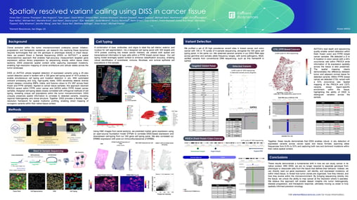
Using a 29-plex targeted variant panel and a 300 gene cell typing panel, we mapped expressed mutations and assigned cell type labels within the same tissue section. This cell typing model incorporates spatial context alongside transcriptional information to improve classification accuracy. In a breast cancer model, this enabled robust identification of endothelial, immune, fibroblast, and luminal epithelial populations and their spatial relationships.
On the variant detection side, the panel covered high-prevalence mutation sites across key cancer-related oncogene genes and was designed to capture critical variant classes, including SNPs, indels, and fusions.
Each of these tells a different biological story. A point mutation in a kinase domain has different implications than a frameshift or a gene fusion and detecting all three within the same spatial framework means researchers can start building a more complete picture of the mutational landscape directly from tissue.
What the data shows
We performed DISS on both FFPE and fresh frozen breast and colon cancer samples and the results demonstrated something important about the nature of tumor heterogeneity. In this experiment, DISS detected expressed variants spanning allele frequencies from ~9-53% and was able to capture both rare and dominant mutations.
Additionally, spatial maps of PIK3CA and CSTL mutations showed regional enrichment of mutations and distinct differences between tumor and adjacent normal tissue visible at the single-cell level.
That kind of regional specificity gets lost when tissue is dissociated. Bulk sequencing can tell you a mutation is present. DISS can show you where it lives.
Validation across orthogonal methods
In the poster, we compared DISS results against independent validation approaches.
Cell typing assignments were compared against expression predictions from STPath, an open-source foundation model that generates marker gene expression estimates. The correlation was strong, giving us confidence that DISS-based cell typing is capturing real biology rather than technical artifact. We also compared ERBB2 expression to post-run immunofluorescence and saw meaningful concordance across the tissue.
For CTSL variant detection, we compared matched bulk RNA-seq findings on same sample and saw comparable results as using DISS. This is a meaningful validation point, particularly for a technology that is detecting mutations directly in tissue without traditional library preparation.
What’s coming next
What we’re sharing now is early data from tissue profiling on AVITI24. The roadmap ahead builds on this and pushes toward the kinds of comprehensive spatial multiomic experiments that have until now required stitching together multiple experiments.
See the complete poster, including variant detection results, spatial maps, cell typing validation data, workflow schematics, and methodology, available to download now.